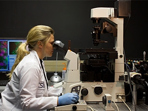
Using a super-microscope built at the Council for Scientific and Industrial Research (CSIR), scientists have made a breakthrough visualisation of a key regulatory protein in body cells.
The CSIR scientists, under the leadership of professor Musa Mhlanga, built the first super-resolution microscope in SA.
In collaboration with colleagues from the University of Cape Town (UCT), the Institut Pasteur and University College London (UCL), the scientists have, for the first time, visualised a key regulatory protein in cells.
This landmark finding appears in the Nature Communications Journal published this month.
According to the CSIR, the protein, called NF-kB Essential Modulator (Nemo), is critical to several basic cellular processes and is implicated in the immune and inflammatory responses, as well as in cancer.
The research institute adds that a mutation in Nemo causes the severe genetic disease incontinentia pigmenti (IP) in females. IP, also known as Bloch-Siemens syndrome, is a genetic disorder affecting the skin, hair, teeth and central nervous system.
Beyond the conventional
Until recently, says the CSIR, there were no simple approaches to visually prove the existence of such highly ordered and organised cytoplasmic structures.
This is largely due to the basic physical properties of light itself, which cannot resolve objects below 200 nanometres in size. Since many structures in the cell may be smaller than this, they cannot be visualised with conventional microscopy, it explains.
"Fortunately, by combining the fields of physics, biology and chemistry, scientists have been able to overcome this problem," says Mhlanga, co-author of the study and technical manager of the Biomedical Translational Research Initiative. The initiative is based at the CSIR and UCT, funded by the Department of Science and Technology (DST).
"So extraordinary was this technological advance that in 2014, a Nobel Prize in Chemistry was awarded for the development of super-resolved fluorescence microscopy."
Professor Ricardo Henriques, a co-author of the paper and research group leader at UCL, says: "Using the state-of-the-art custom-built microscope, we were able to visualise the Nemo structure for the first time, revealing its highly organised and structured nature.
"From the first moment we started imaging Nemo, it was clear that it was forming an organised structure close to the cell surface. This was surprising, as it had never been described before. It was only through the recent advancements in microscopy that we could now observe how Nemo forms higher order structures or lattices."
Eureka!
Dr Fabrice Agou, another co-author on this study, who heads up the Chemogenomic and Biological Screening Platform at the Institut Pasteur in Paris, along with his colleague, professor Alain Isra"el, was among the first researchers to discover and biochemically characterise the Nemo protein.
"Nemo acts like a protein-building block that allows other proteins to build upon it, thus integrating and relaying critical signals within the cells," says Agou.
"Generally, we imagine the cell to be a bag of molecular soup, with an absence of defined structure within the cellular space or cytoplasm. However, through extensive research, it became apparent there was likely to be a special way in which the Nemo protein was structurally assembled in the cytoplasm."
Mhlanga says the work germinated from an insight of Agou and a desire to answer a fundamental, long-standing question regarding signal transduction - the binding of extracellular molecules to receptors that trigger events inside the cell - using cutting-edge technology developed in SA.
Team effort
Co-authors Dr Janine Scholefield and Dr Anca Savulescu - both from the CSIR - were able to interrogate the structure and prove its existence. They also showed that only a few of these supramolecular organising centres were required for a cell to respond to an outside signal.
Says Dr Scholefield: "This study is not only a testament of how the pursuit of basic science can contribute to our greater understanding of how exquisitely our immune system is controlled, but, more importantly, the necessity of international collaborative teamwork for successful scientific research. It also points to the potential to use super-resolution in medical diagnostics."
Mhlanga says such research had a broader significance. "Our goal is that scientists in Africa should not simply be consumers of fundamental scientific discoveries; rather they should be active contributors and producers to this body of knowledge. We would like to train the next generation of scientists in Africa to routinely produce ground-breaking work. In this regard, we are very grateful for the continued support we receive from the DST."
Share